Scientific Calendar August 2024
Post subarachnoid haemorrhagic course
What are typical findings in the cytospin after a subarachnoid haemorrhage?
Red blood cells
Haematoidin crystals in macrophages
Siderophages
Erythrophages
Eosinophils
Congratulations!
That's the correct answer!
Sorry! That´s not completely correct!
Please try again
Sorry! That's not the correct answer!
Please try again
Notice
Please select at least one answer
Scientific Background
Cerebrospinal fluid (CSF)
Cerebrospinal fluid (CSF) is a clear and colourless fluid that surrounds the brain and spinal cord of all vertebrates [1]. Within the central nervous system (CNS), the CSF fills the ventricular system and the subarachnoid space, which is the area between the arachnoid mater and the pia mater.
The CSF facilitates communication between the CNS and the peripheral nervous system, serves as mechanical and immunological protection of brain matter and acts as the transport medium for various substances, such as glucose and immunoglobulins [1]. Examination of the CSF helps the diagnosis of many neurological diseases, such as: CNS infections (e.g. meningitis), inflammatory or autoimmune disorders and neoplasia with possible infiltration into the CNS.
A thorough morphologic analysis of CSF is indicated in every case of elevated CSF cell count to obtain valuable diagnostic information and evaluate therapeutic responses [2,3]. As a rule of thumb, findings of more than five white blood cells (WBC) per µL and any red blood cell (RBC) are considered pathological in adults [3,4].
Subarachnoid haemorrhage (SAH)
A subarachnoid haemorrhage (SAH) is most often caused by a traumatic injury to the brain or by a spontaneous ruptured aneurysm [2]. Risk factors for spontaneous SAH cases include hypertension, smoking and alcoholism [5]. The SAH is accompanied by an inflammatory response, which leads to an increased WBC count [1].
A first sign of bleeding is the detection of erythrophages. After three to four days, these cells exhibit the iron-storage complex haemosiderin in their cytoplasm and are then called siderophages [1]. After around eight days, another product of the haemoglobin degradation process, haematoidin, may be observed. Haematoidin presents itself as yellowish to brown crystals that are either found intra- or extracellularly. Therefore, the presence of siderophages with haemosiderin or haematoidin crystals are indicative for the presence of a SAH and may support in the assignment of time at which the bleeding occurred [1].
Case results
Case and results interpretation
A CSF sample of a male patient in his 70s, who had previously undergone surgery for a SAH, was investigated a week after the removal of the drainage tube. Macroscopically, the CSF sample exhibited a pale orange colour. This is also known as ‘xanthochromia’ and is an indicator for the presence of bilirubin, the haemoglobin degradation product, which occurs after bleeding in the subarachnoid space.
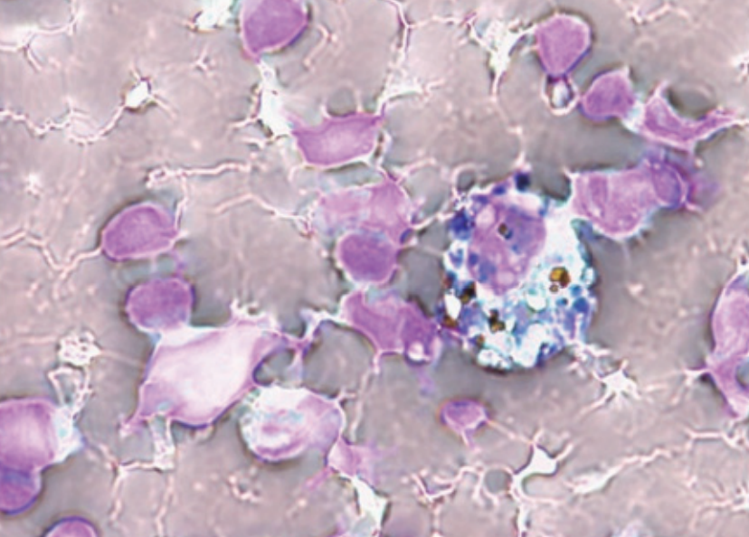
In the cytospin preparation (Fig. 1) stained with May-Grünwald-Giemsa, numerous RBC and siderophages containing basophilic haemosiderin granules and brownish haematoidin crystals were observed.

Scattergram interpretation
In the Body Fluid (BF) mode of the XR analyser, an elevated number of WBC with a predominance of mononucleated cells (MN) were found along with an elevated number of RBC (Table 1).

Table 1 Numerical results of XR BF sample analysis
| WBC-BF | 0.027 × 10⁹/L | |
| RBC-BF | 0.004 × 10¹²/L | |
| MN | 0.023 × 10⁹/L | 85.2 % |
| PMN | 0.004 × 10⁹/L | 14.8 % |
| TC-BF# | 0.027 × 10⁹/L |
| Research parameters | ||
| HF-BF | 0.000 × 10⁹/L | 0.0/100 WBC |
| NE-BF | 0.004 × 10⁹/L | 14.8 % |
| LY-BF | 0.017 × 10⁹/L | 63.0 % |
| MO-BF | 0.006 × 10⁹/L | 22.2 % |
| EO-BF | 0.000 × 10⁹/L | 0.0 % |
The predominance of MN is displayed as green dots in the BF WDF scattergram (Fig. 3). The rightward extension of the MN cluster towards the high SSC area indicates the presence of macrophages with engulfed haematoidin crystals and haemosiderin granules, which were also observed on the cytospin.
Since the patient’s drainage tube was removed one week prior to measurement, the presence of haematoidin crystals was in accordance with the patient’s medical history.
References
[1] Torzewski M et al. (2016): Cerebrospinal fluid cytology: a highly diagnostic method for the detection of diseases of the central nervous system. LaboratoriumsMedizin; 40(s1).
[2] Hrishi A et al. (2019): Cerebrospinal Fluid (CSF) Analysis and Interpretation in Neurocritical Care for Acute Neurological Conditions. Indian J Crit Care Med; 23(2): S115-S119.
[3] Fleming C et al. (2015): Clinical relevance and contemporary methods for counting blood cells in body fluids suspected of inflammatory disease. Clin Chem Lab Med; 53(11):1689.
[4] Jurado R et al. (1990): Cerebrospinal fluid. Clinical methods: the history, physical, and laboratory examinations; 3rd ed. Boston: Butterworths.
[5] Feigin VL et al. (2005): Risk factors for subarachnoid hemorrhage: an updated systematic review of epidemiological studies. Stroke; 36(12): 2773-2780.


