Scientific Image Gallery
Welcome to our Scientific Image Gallery. Here you can find real-life examples of cell images, mostly (but not only) from peripheral blood films, that illustrate typical morphologic characteristics pointing to specific conditions or disorders. This constitutes their diagnostic value.
Click on an image to enlarge it and display a short description.

Leucoerythroblastic condition and schistocytes in a patient with colorectal carcinoma. The occurrence of schistocytes (->) in tumour patients is sometimes an indication of tumour infiltration of the bone marrow.
<p>Leucoerythroblastic condition and schistocytes in a patient with colorectal carcinoma. The occurrence of schistocytes (->) in tumour patients is sometimes an indication of tumour infiltration of the bone marrow.</p>

Cell description:
Size: 10-16 µm
Nucleus: round or slightly indented with condensed and cloddy chromatin, usually invisible nucleolus
Cytoplasm: scanty, weakly basophilic, may have small numbers of azurophilic granules
Morphologically functional subset of lymphocytes cannot clearly be distinguished.
Function: recognize and eliminate threats to the body. Lymphocytes of the innate immune system deliver an immediate response to viral attack. Lymphocytes of the adaptive immune system are specific to a particular antigen.
<p>Cell description: </p> <p>Size: 10-16 µm </p> <p>Nucleus: round or slightly indented with condensed and cloddy chromatin, usually invisible nucleolus </p> <p> Cytoplasm: scanty, weakly basophilic, may have small numbers of azurophilic granules </p> <p>Morphologically functional subset of lymphocytes cannot clearly be distinguished. </p> <p>Function: recognize and eliminate threats to the body. Lymphocytes of the innate immune system deliver an immediate response to viral attack. Lymphocytes of the adaptive immune system are specific to a particular antigen.</p>

In the peripheral blood (May-Grünwald-Giemsa stain) of this patient lymphoma cells can be detected. They show a large, deeply indented nucleus (->) and scanty cytoplasm. Further diagnostics confirmed a mantle cell lymphoma.
<p>In the peripheral blood (May-Grünwald-Giemsa stain) of this patient lymphoma cells can be detected. They show a large, deeply indented nucleus (->) and scanty cytoplasm. Further diagnostics confirmed a mantle cell lymphoma.</p>

Peripheral blood (May-Grünwald-Giemsa stain) of an 82-year old patient with RCMD showing a macrocytic, hyporegenerative anaemia with clear changes of the red blood cells (micro- and macrocytosis, poikilocytosis and some teardrop cells (->)).
<p>Peripheral blood (May-Grünwald-Giemsa stain) of an 82-year old patient with RCMD showing a macrocytic, hyporegenerative anaemia with clear changes of the red blood cells (micro- and macrocytosis, poikilocytosis and some teardrop cells (->)).</p>

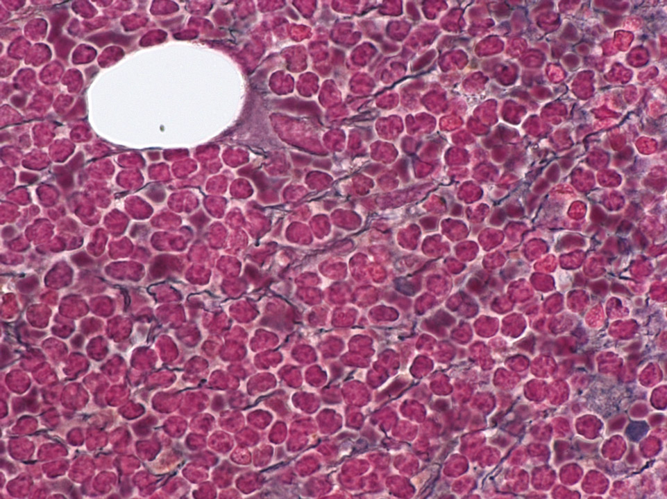
Bone marrow histology (Gomori stain) of a patient showing an infiltration of mantle cell lymphoma cells and an increase in fibres (black lines).
<p>Bone marrow histology (Gomori stain) of a patient showing an infiltration of mantle cell lymphoma cells and an increase in fibres (black lines).</p>

May-Hegglin anomaly belongs to a family of macrothrombocytopenias characterised by mutations in the MYH9 gene. It is a rare autosomal dominant disorder characterised by various degrees of thrombocytopenia that may be associated with purpura and bleeding.
<p>May-Hegglin anomaly belongs to a family of macrothrombocytopenias characterised by mutations in the MYH9 gene. It is a rare autosomal dominant disorder characterised by various degrees of thrombocytopenia that may be associated with purpura and bleeding. </p>


