Scientific Image Gallery
Welcome to our Scientific Image Gallery. Here you can find real-life examples of cell images, mostly (but not only) from peripheral blood films, that illustrate typical morphologic characteristics pointing to specific conditions or disorders. This constitutes their diagnostic value.
Click on an image to enlarge it and display a short description.

Cell description:
Size: 10-12 µm
Nucleus: kidney or U-shaped with clumped chromatin
Cytoplasm: acidophilic
neutrophil: fine reddish granulation
Cell division is not possible anymore and protein synthesis has stopped.
<p>Cell description: </p> <p>Size: 10-12 µm </p> <p>Nucleus: kidney or U-shaped with clumped chromatin </p> <p>Cytoplasm: acidophilic </p> <p>neutrophil: fine reddish granulation </p> <p>Cell division is not possible anymore and protein synthesis has stopped.</p>

Blood film prepared after storage of the EDTA blood for more than one day. A safe morphological differentiation is no longer possible.
<p>Blood film prepared after storage of the EDTA blood for more than one day. A safe morphological differentiation is no longer possible.</p>

Cell description:
Size: 20 µm
Nucleus: kidney- to band-shaped
Cytoplasm: grey and clear with fine azurophilic granules
They are shortly located in the peripheral blood and then move into the tissue where they differentiate into macrophages. Function: Phagocytosis either of harmful pathogens or dead, dying or damaged cells from the blood.
<p>Cell description: </p> <p>Size: 20 µm </p> <p>Nucleus: kidney- to band-shaped </p> <p>Cytoplasm: grey and clear with fine azurophilic granules </p> <p>They are shortly located in the peripheral blood and then move into the tissue where they differentiate into macrophages. Function: Phagocytosis either of harmful pathogens or dead, dying or damaged cells from the blood. </p>
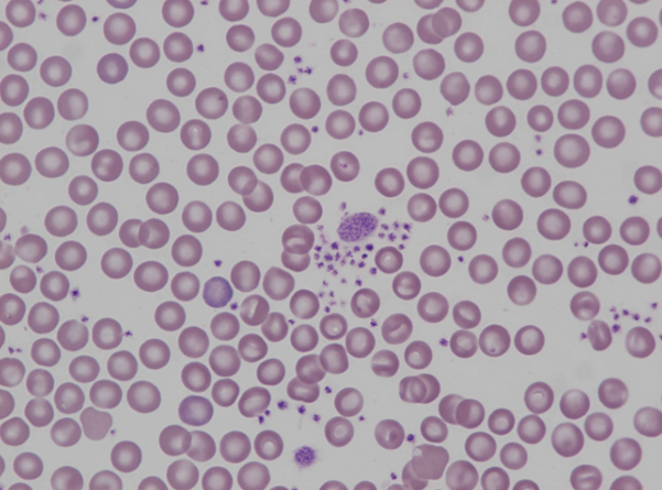
There are two giant platelets surrounded by small platelet aggregates. Giant platelets are occasionally seen in healthy untreated mice. If there is an increased incidence of giant platelets with decreased platelet counts, the bone marrow should be examined for megakaryocytic changes.
<p>There are two giant platelets surrounded by small platelet aggregates. Giant platelets are occasionally seen in healthy untreated mice. If there is an increased incidence of giant platelets with decreased platelet counts, the bone marrow should be examined for megakaryocytic changes.</p>
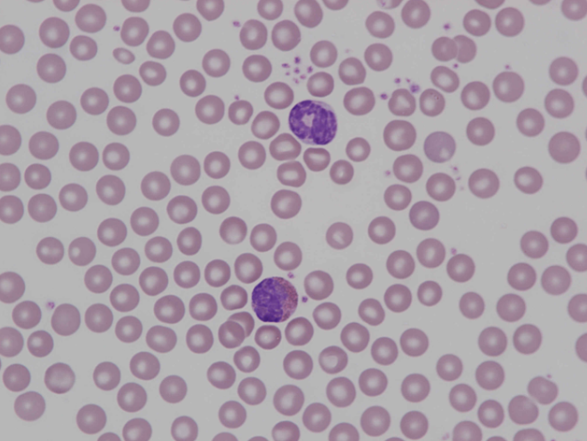
Band neutrophil at the top with a backward-folded appearance of the ring-shaped nucleus. Below that, there is an eosinophil with a nucleus of twisted appearance and numerous round, dark orange stained granules.
<p>Band neutrophil at the top with a backward-folded appearance of the ring-shaped nucleus. Below that, there is an eosinophil with a nucleus of twisted appearance and numerous round, dark orange stained granules.</p>
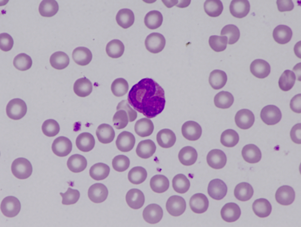
The nucleus of a mouse eosinophil is usually band- or ring-shaped, lighter in colour and slightly more delicate than the nucleus of a neutrophil. The cytoplasm contains large, round, and dark orange stained granules.
<p>The nucleus of a mouse eosinophil is usually band- or ring-shaped, lighter in colour and slightly more delicate than the nucleus of a neutrophil. The cytoplasm contains large, round, and dark orange stained granules.</p>
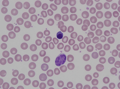
Centrally located erythroblast with a densely stained nucleus and dark purple cytoplasm. The large lymphocyte below that is similar in size to a monocyte but has less cytoplasm.
<p>Centrally located erythroblast with a densely stained nucleus and dark purple cytoplasm. The large lymphocyte below that is similar in size to a monocyte but has less cytoplasm.</p>

