Scientific Image Gallery
Welcome to our Scientific Image Gallery. Here you can find real-life examples of cell images, mostly (but not only) from peripheral blood films, that illustrate typical morphologic characteristics pointing to specific conditions or disorders. This constitutes their diagnostic value.
Click on an image to enlarge it and display a short description.

This patient still has a normal white blood cell concentration of 9,500/μL, but a severely reduced granulocyte count (5%, absolute: 425/μL). Red blood cell count and platelets are inconspicuous. Diagnosis: T-cell large granular lymphocytic (LGL) leukaemia.
<p>This patient still has a normal white blood cell concentration of 9,500/μL, but a severely reduced granulocyte count (5%, absolute: 425/μL). Red blood cell count and platelets are inconspicuous. Diagnosis: T-cell large granular lymphocytic (LGL) leukaemia.</p>

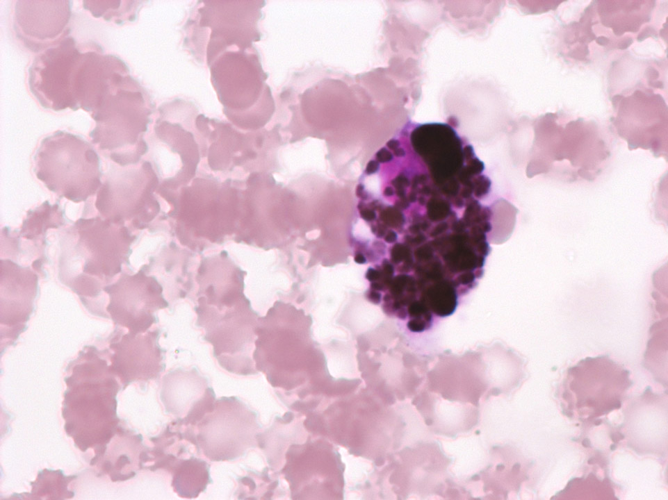
A 'thick blood film' is prepared by spreading a drop of blood on a slide in a way that approximately 20 layers of red blood cells are on top of each other. The slide is then left to dry and subsequently treated with Giemsa solution to lyse the red blood cells. This process leads to a higher density of the malaria pathogens (here: trophozoites of Plasmodium falciparum ->) on the slide so that they can be detected more easily and quickly. The large nuclei are debris from lysed white blood cells.
<p>A 'thick blood film' is prepared by spreading a drop of blood on a slide in a way that approximately 20 layers of red blood cells are on top of each other. The slide is then left to dry and subsequently treated with Giemsa solution to lyse the red blood cells. This process leads to a higher density of the malaria pathogens (here: trophozoites of Plasmodium falciparum ->) on the slide so that they can be detected more easily and quickly. The large nuclei are debris from lysed white blood cells. </p>

Thrombocytopenia and detection of cytoplasm fragments from monoblasts in AML-M5A. Due to their similar size the cytoplasm fragments can produce a falsely elevated platelet count in impedance measurement on certain haematology analysers. Here the initial count was 32,000/μL. The correct platelet value was obtained from the optical channel; it was 7,000/μL. Cytoplasm fragments (C) and a platelet (P) are marked.
<p>Thrombocytopenia and detection of cytoplasm fragments from monoblasts in AML-M5A. Due to their similar size the cytoplasm fragments can produce a falsely elevated platelet count in impedance measurement on certain haematology analysers. Here the initial count was 32,000/μL. The correct platelet value was obtained from the optical channel; it was 7,000/μL. Cytoplasm fragments (C) and a platelet (P) are marked.</p>

Thrombocytopenia (45,000/μL) and platelet anisocytosis as an indication of idiopathic thrombocytopenic purpura (ITP) in a child after viral infection. IPF (immature platelet fraction) in this case was 18.3%.
<p>Thrombocytopenia (45,000/μL) and platelet anisocytosis as an indication of idiopathic thrombocytopenic purpura (ITP) in a child after viral infection. IPF (immature platelet fraction) in this case was 18.3%.</p>

Thrombocytosis, anisocytosis of the platelets and giant platelets in a patient suffering from essential thrombocythaemia (ET). (Also, please note the profound poikilocytosis of the red blood cells.)
<p>Thrombocytosis, anisocytosis of the platelets and giant platelets in a patient suffering from essential thrombocythaemia (ET). (Also, please note the profound poikilocytosis of the red blood cells.)</p>


